Yun-Hee Rhee1,
Seong-Hwa Oh2,
Hyun-Ju Lim3,
Jang-In Shin1,
Chan Kim4,
Chung-Hun Oh1 ![]()
For correspondence:- Chung-Hun Oh Email: choh@dankook.ac.kr Tel:+82-41-550-1918
Received: 18 October 2016 Accepted: 13 January 2017 Published: 26 February 2017
Citation: Rhee Y, Oh S, Lim H, Shin J, Kim C, Oh C. Bone regeneration potential of sub-microfibrous membranes with osteogenic induction of rBMSC for tissue engineering. Trop J Pharm Res 2017; 16(2):271-278 doi: 10.4314/tjpr.v16i2.3
© 2017 The authors.
This is an Open Access article that uses a funding model which does not charge readers or their institutions for access and distributed under the terms of the Creative Commons Attribution License (http://creativecommons.org/licenses/by/4.0) and the Budapest Open Access Initiative (http://www.budapestopenaccessinitiative.org/read), which permit unrestricted use, distribution, and reproduction in any medium, provided the original work is properly credited..
Purpose: To examine the biocompatibility and osteoinductive potential of sub-microfibrous membranes with cells in vitro and in vivo.
Methods: Polylactic acid (PLA) and poly-ε-caprolactone (PCL) were blended at various volume ratios (PLA:PCL = 100:0, 70:30, 50:50, 30:70 and 0:100) and each membrane form was prepared by electrospinning. Cell viability, biocompatibility, and bone regeneration were measured.
Results: The membranes from the PLA/PCL blends prepared by an electrospinning process showed a range of diameter distribution ranging from 1,580 to 550 nm. The cells of 100 % PCL membrane (smallest diameter) exhibited significantly higher adhesion and proliferation than those of the other membranes. Among the membranes from PLA/PCL blends, PCL membrane showed weak inflammatory changes in the early stages of implantation without acute or chronic inflammation. PCL membranes with osteogenically-induced cells successfully stimulated new bone formation in a rate calvarial defect model.
Conclusion: The results indicate that biodegradable PCL sub-microfibrous membrane produced by electrospinning process seems to have excellent biocompatibility, and may be used as a scaffold for bone tissue engineering.
Introduction
Fibrous membranes of biodegradable polymers fabricated by an electrospinning technique have been developed for medical applications such as vascular graft [1], skin substitution [2], nerve regeneration [3], bone tissue engineering [4], and guided bone regeneration for dental surgery [5]. Many studies have been devoted to evaluating the suitability of these applications for in vitro and in vivo models [6]. Biodegradable substance, such as poly (lactic acid) (PLA), Poly-ε-caprolactone (PCL), poly (glycolic acid) (PGA), and their copolymers, are regarded as the ideal materials for medical applications.
To develop an ideal biomaterial, we need to take into account not only its biocompatibility but also the balance between the biodegradation rate of exogenous polymers and the repair rate of the home tissue. Biodegradation rate is stimulated by penetration, attachment, growth, and differentiation of functional cells, but is inhibited by inflammatory responses and tissue rejection reactions. For optimal tissue engineering of damaged tissue, the biodegradation of the implanted scaffold, by host enzymatic and hydrolytic activities, must be followed by tissue replacement and functional restoration such as mechanical strength and original cellular activities.
Some studies have focused on the biodegradability [7], physic-chemical properties [8] and in vitro biocompatibility [2] of aliphatic polyesters and aliphatic polyester blends. In this work, two synthetic polymers were selected; poly-ε-caprolactone (PCL) and poly-lactic acid (PLA). The polymers were blended at different ratios and were subjected to electrospinning. To examine biocompatibility of these membranes, human mesenchymal stem cells (hMSCs) were grown on the PCL/PLA composite membranes, and the tissue responses were examined in the subcutaneous tissues of mice. To assess the efficiency of bone formation, we evaluated the bone hilling activity stimulated by bone marrow-derived stromal cells (BMCs) with or without osteogenic induction on the membranes in a rat calvarial defect model.
Methods
Preparation and characterization of fibrous membrane
PLA (Sigma–Aldrich USA) and PCL (Sigma–Aldrich, USA) were separately dissolved in 2,2,2-trifluoroethanol (TFE, Sigma–Aldrich). The volume ratio of PLA and PCL in the mixed solutions was either 100:0, 70:30, 50:50, 30:70 or 0:100. The mixed solutions were stirred for 24 h. Each solutions were applied into the 10ml syringe to electrospinning. An 15 kV voltage was applied and the tip to collector distance was maintained at 10 cm. The fibrous membranes were dried under vacuum to evaporate the solvent. The morphology of the samples was examined by scanning electron microscopy (SEM), and the diameter of the fibers was calculated from the SEM images.
In vitro viability test
Human mesenchymal stem cells (hMSC) were purchased from Lonza (Walkerville, USA) and maintained in Dulbecco’s modified Eagle’s medium (DMEM) containing 4.5 g/ml D-glucose, 10 % FBS, and 10 U/ml penicillin-streptomycin.
The proliferation of hMSCs on fibrous membranes was evaluated using a MTS assay (Promega, USA). Briefly, hMSCs were seeded at a density of 2 × 10⁵ cells/cm2 on various types of fibrous membranes in a 12-well plate at 0 days. The cells were cultured in the growth medium. Every 24 h, an MTS solution was added to each well, incubated for 30 min, and measured using a microplate reader (Molecular Devices, USA) at 450 nm. The proliferation of hMSCs was observed by confocal microscopy. The cells were processed, as described in the MTS assay. All membranes were fixed with chilled ethanol every 24 h, and 50 μg/ml propidium iodide solution was added to each well. The membranes with the cells were incubated for 15 min and washed with PBS briefly. Each time the number of cells in the fibrous membranes was counted and photographed by LSM510 confocal microscopy (Carl Zeiss, Switzerland).
In vivo biocompatibility test
The animal protocol used in this study was reviewed and approved based on ethical procedures and scientific care by the Dankook University-Institutional Animal Care and Use Committee (DKU-IACUC). To implant the fibrous membranes, incisions were cut on the dorsal section of each mouse and the fibrous membranes were and implanted into the incisions. The incisions were then sutured. After two weeks, the mice were sacrificed, and the entire graft was harvested with surrounding tissue. The samples were fixed with 4 % paraformaldehyde and were subjected to Hematoxylin-Eosin staining.
Assessment of bone regeneration in calvarial defects
To evaluate whether bone marrow-derived stromal cells (BMCs) with osteoinduction participate in in vivo bone formation, a calvarial bone defect model was used for this assay in 8-week-old Sprague-Dawley (SD) rats. BMCs were induced in osteogenic media for 7 days after growth in DMEM for 7 days. The osteogenic media consisted of 50 mM L-ascorbic 2-phosphate, 10 mM glycerol-2-phosphate disodium, and 1 μM dexamethasone, and the medium was replaced every 3 days.
Under general anesthesia, induced by an intramuscular injection of ketamine/rumpun (80 and 10 mg/kg), the external cortical plates were removed using a 5-mm trephine bur with saline irrigation [9] [10]. One asymmetrical defect was made on the right side of the midline of each rat. Extreme care was taken not to harm the brain membranes. The bone defects were washed with normal saline, and membranes were then applied to the defects.
Animals were divided into three groups: Group 1, PCL membrane only (n=3); Group 2, PCL membrane with non-induced rBMCs (n=3); Group 3, PCL membrane with osteogenic induction of rBMCs (n=3); No immunosuppressants were used. After 4 weeks, the rats were euthanized, and the transplanted constructs were dissected carefully and taken out of the surrounding soft tissue.
Microradiographs of the skull were performed using an Image Station FX (Kodak, Rochester, NY) for 6 s at 12.5 kVp. After the radiographs had been taken, the defected calvaria samples were de-calcified with 20 % EDTA. The samples were then dehydrated in a graded series of ethanol and embedded in paraffin. The center of the defect area was sectioned to a 4 μm thickness, stained with Masson’s Trichrome, and photographed using an optical microscope (Olympus BX51, Olympus, Miami, FL, USA).
Statistical analysis
All results are presented as mean and stabdard deviation (SD) and were evaluated using one-way analysis of variation (ANOVA) and also Tukey’s analysis for pair wise comparison. Differences were considered significant at p < 0.05.
Results
Structural morphology of the fibrous membrane
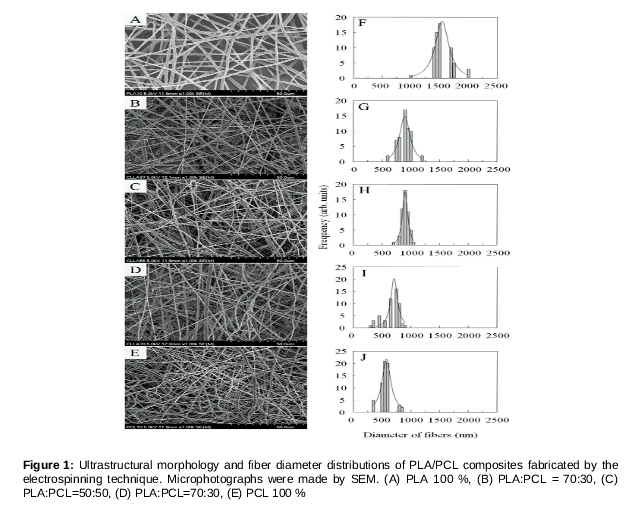
As shown in , fibrous membranes produced from different volume ratios of PLA and PCL by an electrospinning technique led to a range of diameter distributions. The three-dimensional fibrous structures with polygonal, interconnected pores and randomly oriented fibers were evaluated for their diameter range. The diameters of homogenous PLA fabrications was determined to be 1,580 ± 232 nm (A and F), PLA: PCL=3:7 835 ± 123 nm (B and G), PLA: PCL = 5:5 842±97 nm (C and H), PLA: PCL = 3:7 743 ± 173 nm (D and I), and the homogenous PCL 558 ± 142 nm (E and J).
Viability of hMSC on the fibrous membrane
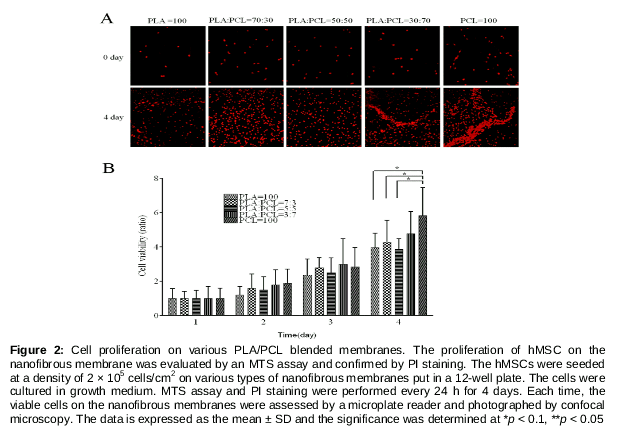
The viability of the hMSCs on the fibrous membrane was examined using an MTS assay and PI staining (). On day 0, the hMSCs were seeded onto the various types of membrane, and their attachment and proliferation were observed after 7 days. As shown in a, the hMSCs were showed exponential growth patterns, without any cytotoxicity, for all types of membranes until day 4. Cells on PCL fibrous membranes showed a significantly higher viability than those on any of the other materials, and were in a relatively quiescent state throughout the 7 day period after seeding, compared to those of other membranes.
In vivo biocompatibility
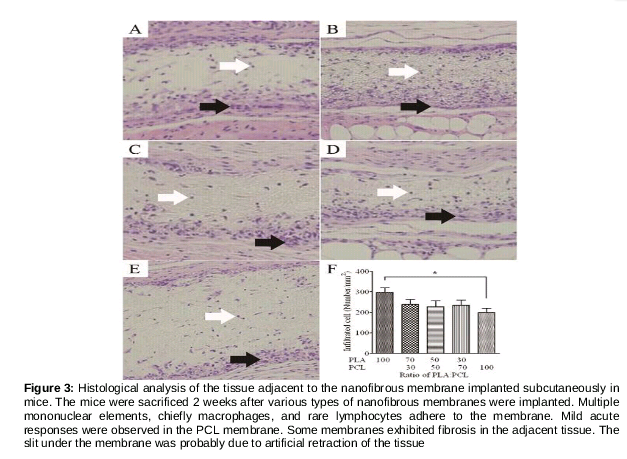
Histological analysis by optical pathological microscopy was performed to make a comparison of the infiltration to various types of membrane. The pore size of the fibrous membranes is too small for cells to penetrate in vitro. However in vivo cellular infiltration was observed in the subcutaneous tissue of mice examined in week 2. As shown in , histological examination of subcutaneous implantation with fibrous membranes revealed an infiltration of histiocytes and lymphocytes in the area of the margin and center, respectively. There was no evidence of acute or chronic inflammation in any groups. Our results showed that hypertrophy of lining histiocytes and infiltration of lymphocytes in the membranes gradually decreased with increasing ratio of PCL. However, the difference was not determined to be statistically significant (p > 0.5). For this reason, PCL seems to be a suitable candidate as a degradable fibrous membrane for use in biomaterials.
Bone healing in rat calvarial defect model
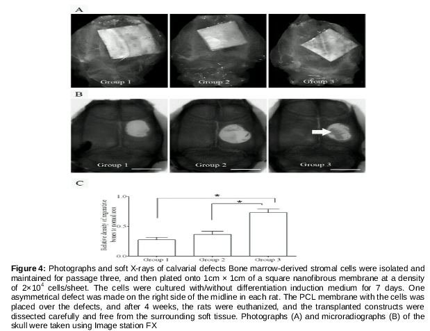
Bone regeneration effects were evaluated in the rat calvarial defect model by Soft x-ray and histological staining after four weeks. From the Soft x-ray observation (B, implantation of fibrous membranes with non-induced rBMSC (group 2) to calvarial defected rats showed limited bone formation at the periphery, as did membrane only group (group 1). In contrast, implantation of the membrane with osteogenic induction of rBMSC (group 3) showed a significantly greater extent of bone healing, both on the inside and peripheral regions of the defect, compared to other groups. The quantitative analysis of bone density was consistent with Soft X-ray images. The relative bone regeneration area of group 3 was 0.73 ± 0.08 mm, which was significantly greater than that of group 2 (0.36 ± 0.07 mm) and group 1 (0.28 ± 0.03 mm).
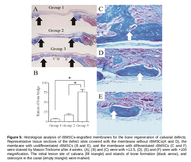
Histological images of the implanted membranes 4 weeks after surgery are shown in . Masson’s Trichrome (MT) stained sections were analyzed with light microscopy at various levels of magnification, yielding information on the quality and histologic characteristics of bone regeneration. MT-stained Histological sections confirmed a significant amount of bone healing, consistent with the island of new bone in group 3 ( A), and also revealed a large number of collagen tissues near the island formation of new bone tissue. All of the newly formed bones tissues had osteocytes in the canal cavity ( C, D, E).
In quantitative analysis (B), the average of bony bridging ratios in the calvarial defects was 12.3 % for PCL membranes only (Group 1), 22.2 % for PCL membranes with non-induced rBMSC (Group 2), and 73.1 % for PCL membranes with osteogenic induction of rBMSC (Group 3; A, B). The bone bridging ratios histologically correspond well to Soft x-ray data obtained.
Discussion
The fiber diameter, biocompatibility, and degradation rate of the blended polymers are essential for guided bone regeneration in dental surgery. This provides vital information regarding protection from soft tissue penetration during the hard tissue regeneration. Several observations have shown that pure PLA is hard and brittle and that adding PCL can change its original properties to make it softer and more flexible. The elongation properties of PLA increase with the addition of PCL, but the strength decreased as the elongation properties increased [8]. PLA can degrade more quickly than PCL, and so, it needs a shorter time to fully regenerate tissues of damaged organs. In other words, the addition of PCL to membranes hastens regeneration.
The fiber diameter of the blended polymers was estimated by SEM images. As shown in , fiber diameters of the blended polymers ranged from 500 nm to 1.5 μm. The relative fiber diameters of the blended polymers were ranked as following: PLA > PLA/PCL (7/3) > PLA/PCL (5/5) > PLA/PCL (3/7) > PCL. Many studies have reported that fibrous membranes blended with different biodegradable polymers by an electrospinning technique resulted in a range of diameter distributions from 10 nm to a few micrometer [11,12].
Biocompatibility was addressed regarding in vitro viability and in vivo tissue response. From our results, viable cells were more abundant on the PCL fibrous membrane than other PLA/PCL blended membranes, and the dominant diameter of the PCL fibrous membranes are the smallest among the PLA/PCL blended membranes. The fiber diameter of the fibrous membranes affects cell proliferation, migration, and penetration in the membranes. The rat hippocampal astrocyte (HA) and rat cerebro-microvascular endothelial cells (CEC) could penetrate to PCL microfibrous membranes, but not to PCL sub-microfibrous membranes [13]. For the viability of HUVECs, PCL membranes with a fiber diameter of 1.16 ± 0.17 μm were more suitable than ones with fiber dimensions of 7.02 ± 1.03 μm [14].
Several works demonstrated that electrospun fibrous membranes showed a minimized foreign body reaction in animal models in vivo, such as rat [15], rabbit [16] and sheep [17]. From our results, all membranes made of the blended polymers showed mild foreign body reaction, and the membrane with PCL had a lower inflammation than others, due to the less acidic products of degradation.
Recently, several studies focused on stem cell transplantation with several osteogenic factors, such as BMP-2 [18], fibroblast growth factor 2 [19], and the demineralized bone matrix [20], for bone regeneration in calvarial defect animal models. Moreover, multipotent adult stem cells seeded into the polyglycolic acid mesh can stimulate bone regeneration after 12 weeks, and without any osteogenic factors, in a rat calvarial defect model [21].
In the bone repairing animal model, transplantation with differentiated stem cells showed a better result than that with the control or undifferentiated stem cells [22,23]. These results are in agreement with our observation that the PCL fibrous membrane with osteogenic induction of rBMCs in the calvarial defect model stimulated a more compact bone regeneration than the one with non-induced rBMCs after 4 weeks. Furthermore, bone healing was observed in MT staining, which revealed areas of cortical bone with osteocytes.
Conclusion
The findings of the present study demonstrate that 100 % PCL fibrous membrane shows better biocompatibility and viability than PLA blended forms, and that bone regeneration ability is improved on PCL sub-microfibrous membranes with osteogenic-induced cells, compared to that with non-induced cells in a calvarial defect model.
Declarations
Acknowledgement
References
Archives


News Updates